Department of Surgery Jars
General principles
What we usually refer to as Jars examination is, in reality, a surgical pathology one. This realization is important as it has its bearing on your description of specimens and on the subsequent questions and answers.
The usual scenario
Hold the jar and have a thorough look at its all aspects (4 sides, roof and bottom).
Describe the contained specimen in a systematic manner,
e.g., .shape, size, colour, and surface. If it is a hollow organ describe its inner surface, wall and outer surface. The presence of a ligature or a cut in the specimen should also be mentioned.
If you know the organ name it, but you may be asked how you recognized it. This will be mentioned later in relation to each organ in relevant sections.
If you cannot recognize the origin of this specimen describe it in detail to reach the nearest possibility.
The key points in recognition are:
Whether it is a hollow or a solid organ.
Shape.
Attachment to a pedicle, a tubular structure (ureter), or mesentery.
Describe the lesion , i.e., the abnormality.
o This might be a swelling or an ulcer, whereby you will follow the scheme of describing such lesions by inspection in a living patient, e.g., number, site, size, edge, surface, ��etc.
o The abnormality might be a mere enlargement of the organ, thickened wall, dilated lumen, â?¦etc.
Diagnosis . For most specimens, and based on the findings, you will be requested to name an anatomical and a pathological diagnosis. For example the diagnosis might be ileo-caecal intussusception. A pathological diagnosis also includes complications, and in our example it might be intestinal gangrene.
Discussion
o As this is a surgical pathology examination, you are likely to be asked about the histological picture, the possible functional abnormalities, and the possible complications.
o Other questions may include clinical picture, diagnosis and treatment.
|
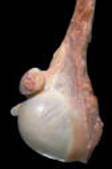
Gall bladder |
|
|
Pear shaped Hollow organ Blind broad end and open narrow end that may be tied by a ligature May contain stones |
|
|
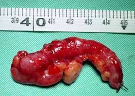
Appendix |
|
|
Hollow tubular organ Usual length 5-10cm Blind end and an open end that may be tied by a ligature Has a mesentery (mesoappendix) |
|
|
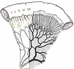
Small intestine |
|
|
Hollow tubular structure Two open ends Mesentery |
|
|
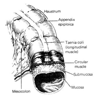
Large intestine |
|
|
Hollow tubular structure Two open ends May have a mesentery (transverse and sigmoid colons) Taenia coli on outer surface (longitudinal muscle bundles) Haustrations and appendices epiplocae may be seen |
|
|
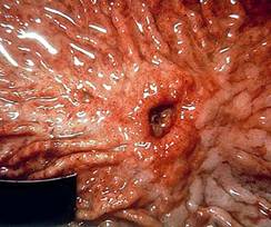
Stomach |
|
|
Wide capacious hollow viscus Characteristic mucosal folds (rugae) Inner surface may contain an ulcer(s) or a tumour |
|
|
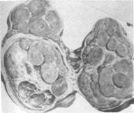
Thyroid |
|
|
Butterfly-shaped when the specimen is that of total or subtotal thyroidectomy Solid organ |
|
|
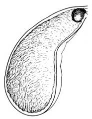
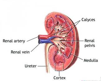
Kidney |
|
|
Bean-shaped Solid organ. In case of an advanced hydronephrosis the kidney is transformed into a bag of urine Has a hilum with vessels and ureter May be bisected to show its collecting system of calyces and pelvis |
|
|
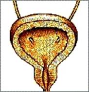
Urinary bladder |
|
|
Hollow muscular organ Two ureters attached Bladder neck Prostate may be included in the specimen |
|
|
Testis |
|
|
Solid organ Globular Attached pedicle (vas deferens) |
|