
Department of Surgery Jars
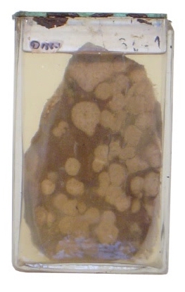
| Description | Multiple Liver Metastases |
| Author | Department of Surgery |
| Copyright | Cairo University - Faculty of Medicine |
Description
This jar contains a slice of the liver that contains multiple grayish white solid lesions. They mostly have irregular outline and range in size from 1 to 3 cm in diameter. Those that reach the surface show a central depression (umbilication).
Diagnosis
Multiple liver metastases
Possible questions
Q. What is the commonest malignant liver tumour?
A. Metastases
Q. Why is it diagnosed as secondaries and not a primary tumour?
A. A primary liver tumour is usually solitary and usually occurs on top of liver cirrhosis. Multiplicity, whether it is in the liver, lungs, brain or bone, usually favours secondaries.
Q. What is the usual origin?
A. Colorectal cancer, stomach cancer, pancreatic cancer (spread through the portal vein) and breast cancer.
Q. What is the microscopic picture
A. Adenocarcinoma
Q. What are the clinical features?
Q. What is the tumour marker that is most likely to be elevated?
A. Carcinoembryonic antigen (CEA). In cas of hepatocellular carcinoma it is alpha fetoprotein that is elevated.