
Department of Surgery Jars
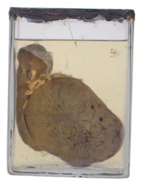
| Description | Renal Cell Carcinoma (Hypernephroma) |
| Author | Department of Surgery |
| Copyright | Cairo University - Faculty of Medicine |
Description
This is a jar that contains one half of a longitudinally bisected kidney. The lower pole is occupied by a solitary ovoid mass that measures about 13 X 9 cm in maximum diameters. The mass grossly looks well-localized and does not invade the outer surface of the kidney. It, however, compresses the pelvicalyceal system. The cut section shows areas of haemorrhage and necrosis.
Diagnosis
Renal cell carcinoma (hypernephroma)
Possible questions
Q. What is the histological picture?
Q. What are the possible clinical features?
A. Classic features include in descending order, haematuria (has its specific features), pain and a loin mass.
Rare features that are related to complications and to the production of hormones.
Q. What are the possible complications and methods of spread?
Q. What is the treatment.
Remember that renal cell carcinoma looks grossly as if it were well-localized, but in fact it shows microscopic invasion of the collecting system that causes haematuria and of the veins in the form of tongue of tissue.