
Department of Surgery Jars
| Description | Osteosarcoma of Upper Humerus. |
| Author | Department of Surgery |
| Copyright | Cairo University - Faculty of Medicine |
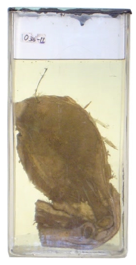
Description
This is a jar that contains one half of a
vertically-bisected arm, elbow and upper forearm of a child. This is
known by the presence of epiphyseal cartilages. There is a
bone-destructive tumour that arises from the metaphysic of the upper
humerus and measures about 12 X 12.cm. The tumour destroyed bone
architecture in upper humerus and extended in the surrounding soft
tissue. It, however, does not invade the epiphysis. The cut section
shows colour heterogenicity with dark areas that are suggestive of
haemorrhage and necrosis.
Diagnosis
Osteosarcoma of upper humerus
Possible questions
Q. What is the usually affected age group?
A. Children.
Q. What are the common sites of
origin of osteosarcoma?
A. Metaphyses of growing ends of bone. In the upper limb this means
upper humerus and lower radius and ulna. In the lower limb it means the
lower end of femur and upper ends of tibia and fibula. The commonest
site, however, is the lower end of femur.
Q. What are the histological
features?
Q. What are the methods of spread?
Q. What are the radiological features?
Q. What is the treatment?