
| Description | Renal Cell Carcinoma (Hypernephroma) |
| Author | Department of Pathology |
| Copyright | Cairo University - Faculty of Medicine |
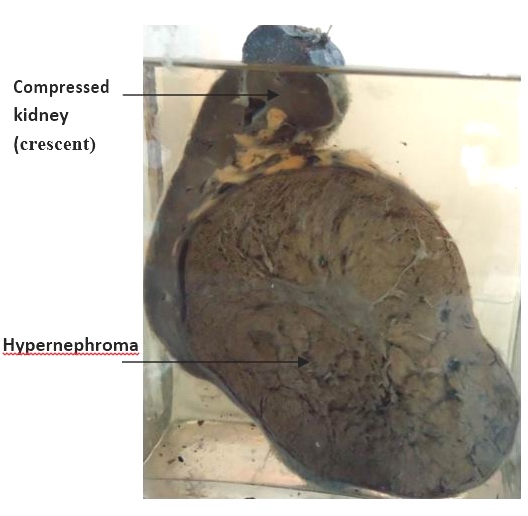
Specimen:
Half of kidney
Gross Pathology :
1. The kidney is enlarged.
2. Cut section shows large oval mass, 15X9 cm arising from the lower pole of the kidney.
3. The mass is non-capsulated but well defined with a yellowish cut surface showing areas of necrosis and hemorrhage.
4. The rest of the kidney is compressed at the upper pole and appears as a crescent.
Diagnosis :
Renal cell carcinoma (hypernephroma).