
| Description | Chronic Fibrocaseous Pulmonary Tuberculosis Associated with Tuberculous Pneumonia |
| Author | Department of Pathology |
| Copyright | Cairo University - Faculty of Medicine |
Specimen:
Sectioned lung
Gross Pathology:
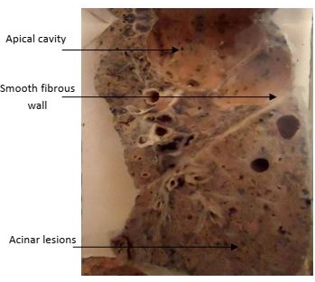
1. A large apical cavity (9 cm in diameter) is seen with a fibrotic wall, and yellow caseous lining traversed by ridges (representing thickened bronchi and blood vessels).
2. The lower lobe shows multiple yellow caseous foci (acinar lesions).
3. The foci at the base get fused (tuberculous pneumonia).
4. The covering pleura shows greyish white fibrous thickening and fibrous adhesions.
5. The tracheobronchial lymph nodes show minimal tuberculous lesions and anthracosis.
Diagnosis:
1. Chronic fibrocaseous pulmonary tuberculosis associated with confluent tuberculous pneumonia.
2. Pleural fibrosis and adhesions.