
| Description | Septic Bronchiopneumonia Associated with Fibrinous Pleurisy |
| Author | Department of Pathology |
| Copyright | Cairo University - Faculty of Medicine |
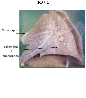
Specimen:
Sectioned left lung of a child.
Gross Pathology :
1. The cut surface of the lung shows multiple small suppurative foci, some showing the cross section of bronchi
3. The lung tissue between the foci appear dark hyperemic.
4. The covering pleura is dull, opaque and greyish due to fibrin deposition
Diagnosis:
1. Bronchopneumonia.
2. Fibrinous pleurisy.