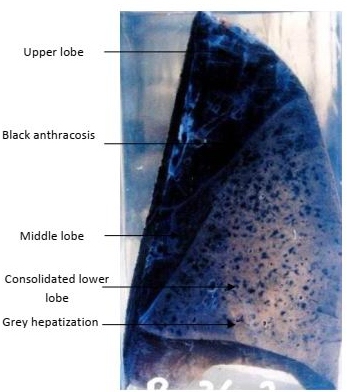

| Description | Lobar Pneumonia, Grey Hepatization of the Right Lower Lobe. |
| Author | Department of Pathology |
| Copyright | Cairo University - Faculty of Medicine |

Specimen:
Section in the right lung.
Gross Pathology :
1. The lower lobe is grayish in color, swollen and consolidated.
2. The cut margins are sharp denoting firm consistency.
3. The covering pleura is dull, opaque and greyish due to fibrin deposition.
4. The upper and middle lobes are collapsed with scattered black anthracotic spots.
Diagnosis:
1. Lobar pneumonia, grey hepatization of the right lower lobe.
2. Fibrinous pleurisy.
3. Anthracosis.