
| Description | Lobar Pneumonia of Left Upper Lobe Associated with Abscess Formation and Fibrinous Pleurisy |
| Author | Department of Pathology |
| Copyright | Cairo University - Faculty of Medicine |
Specimen:
Sectioned left lung (bisected).
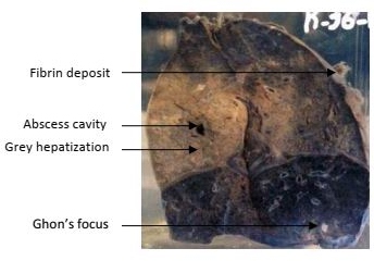
Gross Pathology :
The upper lobe is grayish in color, swollen and consolidated.
The cut margins are sharp denoting firm consistency.
A small abscess cavity (1 cm) is seen.
The covering pleura is dull, opaque and greyish due to fibrin deposition.
The lower lobe shows a small calcified focus (Gohn’s focus).
Diagnosis:
1. Lobar pneumonia of left upper lobe with abscess formation.
2. Fibrinous pleurisy.
3. Ghon’s focus of the lower lobe.