
| Author | Pathology Department |
| Copyright | Cairo University- Faculty of Medicine |

Specimen:
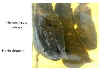
Multiple lung slices
Gross Pathology :
1. The lung slices shows multiple hemorrhagic infarcts.
2. The infarcts are pyramidal in shape, with their bases towards the surface.
3. The covering pleura is dull, opaque and greyish due to fibrin deposition.
Diagnosis:
1. Multiple hemorrhagic lung infarcts.
Fibrinous pleurisy