
| Description | Massive Cerebral Hemorrhage |
| Author | Department of Pathology |
| Copyright | Cairo University - Faculty of Medicine |
Specimen:
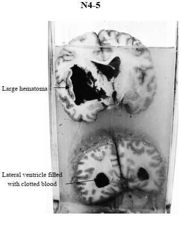
Two coronal sections of the brain.
Gross Pathology:
1. The right hemisphere is slightly swollen
2. The right basal ganglia are destroyed and replaced by large brown hematoma.
3. The lateral ventricles are filled with brown clotted blood.
4. The aqueduct is blocked by clotted blood
5. The basilar artery shows yellow raised nodules of atherosclerosis.
Diagnosis:
1. Massive cerebral hemorrhage.
2. Cerebral atherosclerosis.