
| Description | Craniopharyngioma. |
| Author | Department of Pathology |
| Copyright | Cairo University - Faculty of Medicine |
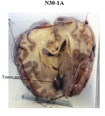
Specimen:
Coronal section of the brain of a child.
Gross Pathology:
1. Cut section shows an oval tumor mass, 5X2 cm, replacing the hypothalamus and protruding into the third ventricle.
2. The distal part of the mass is solid with whitish granular calcifications.
3. The proximal part is cystic with brown coagulated material.
Diagnosis:
Craniopharyngioma.