
Specimen:
Sectioned uterus.
Gross Pathology:
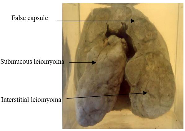
1. The uterus is asymmetrically enlarged.
2. The uterine wall is thickened and shows one submucous and multiple variable-sized interstitial rounded well-defined tumor masses.
3. The masses show greyish white whorly cut section. They are surrounded by compressed uterine muscles forming pseudo-capsule.
Diagnosis
Multiple uterine leiomyomata (fibroids) of the interstitial and submucous types.