
| Description | Cavernous Hemangioma of the Liver. |
| Author | Department of Pathology |
| Copyright | Cairo University - Faculty of Medicine |
Specimen:
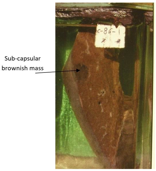
A slice of liver
Gross Pathology :
1. The cut section of the liver shows a sub- capsular well defined non-capsulated dark brown mass measures about 2 cm in diameter.
2. Cut section of the mass is spongy and brownish (multiple vascular spaces filled with clotted blood, separated by fibrous septa).
Diagnosis:
Cavernous haemangioma of the liver.