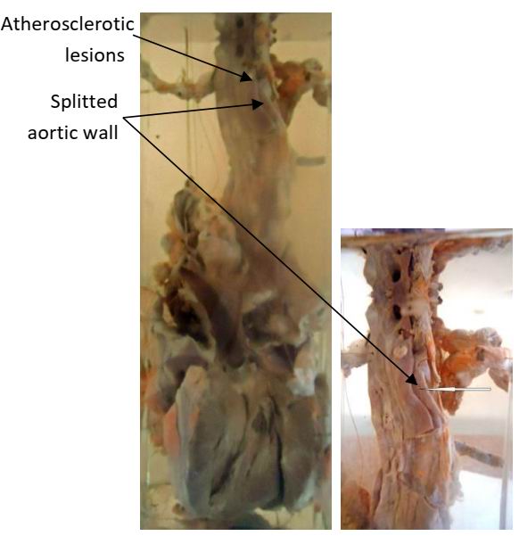

| Description | Aortic Atherosclerosis |
| Author | Department of Pathology |
| Copyright | Cairo University - Faculty of Medicine |

Specimen:
Heart with open left ventricle and part of thoracic aorta.
Gross Pathology:
1. The intima of the aorta shows raised yellow atherosclerotic patches.
2. The wall of aorta is split into two layers (dissecting aneurysm).
Clotted blood is seen between these two layers.
3. The left ventricle is hypertrophied.
Diagnosis:
1. Aortic atherosclerosis.
2. Dissecting aneurysm of aorta.
3. Left ventricular hypertrophy.