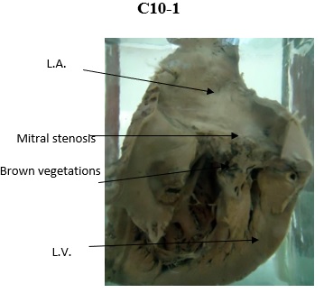

| Description | Rheumatic Endocarditis of the Mitral Valve with Stenosis. |
| Author | Department of Pathology |
| Copyright | Cairo University - Faculty of Medicine |

Specimen:
Opened heart.
Gross Pathology:
1. Mitral valve shows thick grayish white opaque fused cusps with stenosed orifice.
2. The chordae tendineae are shortened and the papillary muscles are hypertrophied.
3. Large brownish vegetations on both surfaces of the cusp.
4. The left atrium is hypertrophied and dilated. Its posterior wall shows an opaque whitish patch (MacCallum’s patch).
Diagnosis:
1. Chronic Rheumatic Endocarditis of the mitral valve with stenosis.
2. Subacute infective endocarditis of the mitral valve.
3. Hypertrophy and dilatation of the left atrium.